Bone Cross Section Histology / Bone Histology - Bone - histology slide : Bone decalcification is the removal of the mineral component using an acid, leaving the bone soft and easy to cut.
Bone Cross Section Histology / Bone Histology - Bone - histology slide : Bone decalcification is the removal of the mineral component using an acid, leaving the bone soft and easy to cut.. A cross section of a human long bone. Jump to navigation jump to search. The section may be either cross section (x.s.) or longitudinal section (l.s.). (a) midshaft cross section of the femur in normal light. Note the large nutrient canal and the single growth mark (arrow).
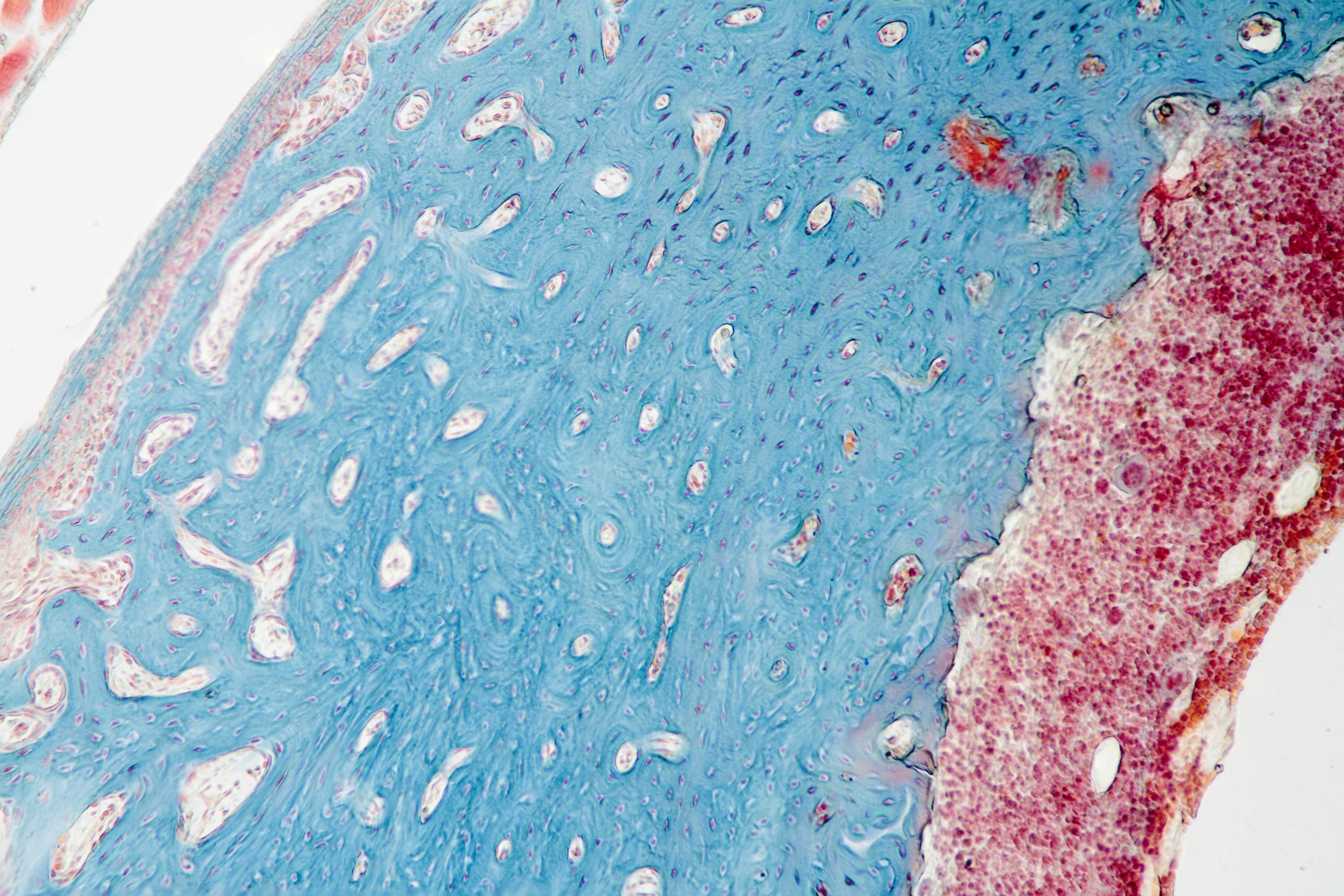
At the outer regions of the section, you can see a dense, thick layer of compact bone. Available at the itunes store and for android users at the google play store. The term 'bone marrow' (bm) refers to the tissue occupying the cavities under the cortex within the this chapter will describe the histology of bm in the trephine biopsy. Anyway, examine the fibers cut in xs to see that the nuclei are located in the center of the fibers (you may need to use oil emersion). A cross section of any bone will demonstrate these two types of bones.
Free photo: Femur Bone Cross Section - Aged, Medicine ... from jooinn.com Dry bone is cut and polished before mounting on a slide. Histology of bone gross structure • the diaphysis is the shaft and notably comprises the marrow cavity. Learn vocabulary, terms and more with flashcards, games and other study tools. Cross section of a long bone. From wikimedia commons, the free media repository. White dashed rectangles indicate areas of image magnification. What follows is primarily a guide to observing particular features microscopically. This is a cross section through decalcified bone.
Muscle attachments are visible along the outer surface.
In addition to discussing the cellular constituents of bone and the architectural arrangement of their products. From wikimedia commons, the free media repository. This is a cross section through decalcified bone. The inner portion of the bone is composed of trabecular bone and the intervening bone marrow. Anyway, examine the fibers cut in xs to see that the nuclei are located in the center of the fibers (you may need to use oil emersion). Histology hint from sarah bellham: White dashed rectangles indicate areas of image magnification. The section may be either cross section (x.s.) or longitudinal section (l.s.). The term 'bone marrow' (bm) refers to the tissue occupying the cavities under the cortex within the this chapter will describe the histology of bm in the trephine biopsy. Note the large nutrient canal and the single growth mark (arrow). Learning objectives describe the histology of bone tissue compare and contrast compact and spongy bone most bones contain compact and spongy osseous tissue, but their distribution and concentration. Accuracy of the tested digitization method was expressed by. Bones protect the various organs of the body, produce red and white blood cells, store minerals.
The inner portion of the bone is composed of trabecular bone and the intervening bone marrow. White dashed rectangles indicate areas of image magnification. Jump to navigation jump to search. First, study cross sections (slides 51 and 93b). Of the four basic tissue types (epithelium, connective tissue, muscle and nervous tissue), connective tissue is the most diverse.
Bone Cross Section Diagram Card | Zazzle from rlv.zcache.com Use the illustrations in your textbook as a guide and identify with the scanning objective the following structures. The functions of bone tissue in histology are defined as coinciding with the functions of all skeletal connective tissue, but this tissue has a number of unique properties. • now, let's point out these histological structures in bone specimens. A cross section of any bone will demonstrate these two types of bones. There are two ways to study bone histology. Bone histology of fossil tetrapods: *blood vessels *nerves *loose connective tissue. This is an online quiz called bone histology bone cross section.
White dashed rectangles indicate areas of image magnification.
Learning objectives describe the histology of bone tissue compare and contrast compact and spongy bone most bones contain compact and spongy osseous tissue, but their distribution and concentration. 'compact or cortical bone is usually thick dense bone that forms the outer shell cross sections of the bone when studied under the microscope reveal quite a different picture. Keep in mind that the word bone can refer to either a type of tissue or to the organ. Advancing methods, analysis, and interpretation. Accuracy of the tested digitization method was expressed by. Use the illustrations in your textbook as a guide and identify with the scanning objective the following structures. Is continuous throughout life and involves a combination of bone synthesis and removal. At the outer regions of the section, you can see a dense, thick layer of compact bone. The functions of bone tissue in histology are defined as coinciding with the functions of all skeletal connective tissue, but this tissue has a number of unique properties. Bone decalcification is the removal of the mineral component using an acid, leaving the bone soft and easy to cut. The term 'bone marrow' (bm) refers to the tissue occupying the cavities under the cortex within the this chapter will describe the histology of bm in the trephine biopsy. There is a printable worksheet available for download here so you can take the quiz with pen and paper. A cross section of a typical osteon or haversian system.
Accuracy of the tested digitization method was expressed by. Histology hint from sarah bellham: The histology of compact bone. Of the four basic tissue types (epithelium, connective tissue, muscle and nervous tissue), connective tissue is the most diverse. This is an online quiz called bone histology bone cross section.
Histology Quiz #1- Bone and Cartilage at Hamilton College ... from classconnection.s3.amazonaws.com Histology classification of bone tissue. Both sections have been decalcified in order to make it easier to cut the bone into thin sections, but the collagen is still present in the slides. Cross section of a long bone. In addition to discussing the cellular constituents of bone and the architectural arrangement of their products. Dry bone is cut and polished before mounting on a slide. White dashed rectangles indicate areas of image magnification. Is continuous throughout life and involves a combination of bone synthesis and removal. Contents (click on desired chapter).
The histology of compact bone.
There is a printable worksheet available for download here so you can take the quiz with pen and paper. First, let's look at a section of compact bone. Cross and longitudinal sections (unstained). Note the large nutrient canal and the single growth mark (arrow). What follows is primarily a guide to observing particular features microscopically. In these sections, the trapped air bends the light giving a dark image; Learn vocabulary, terms and more with flashcards, games and other study tools. Histology classification of bone tissue. A cross section of a human long bone. Accuracy of the tested digitization method was expressed by. As noted in the main text, it was puzzling that the uniqueness of plesiosaurian bone histology was not recognized before, despite the long history of. Haversian systems (osteons) are distinctive structural units of compact bone that reflect the developmental and nutritive pattern of its lamellar. In development there are 2 separate signaling pathways for pattern formation and the formation of bone itself.
Preparation and sectioning of specimens bone cross section. Bone decalcification is the removal of the mineral component using an acid, leaving the bone soft and easy to cut.


0 Komentar